Knee Muscle Anatomy Mri / Mri Knee Joint Anatomy - Check the positioning block in the other two planes.. When interpreting the proton density images it. Anatomy arthrogram anatomy basic shoulder mri. Stanford msk mri atlas has served over 1,000,000 pages to users in over 100 countries. This long muscle flexes the knee. Anatomy basic knee mri checklist.
Please email baodo at stanford.edu Atlas of knee mri anatomy. Mr imaging of knees having isolated and combined ligament injuries. Stanford bone tumor ddx | iss/ssr msk lectures | search ocad cases | stanford virtual readouts stanford msk mri atlas has served over 1,000,000 pages to users in over 100 countries. Both the pronounced accuracy of the mri and the high prevalence of knee disorders, makes the knee mri the most frequently ordered imaging procedure of the.
The Knee Musculoskeletal Key from musculoskeletalkey.com Abnormal anatomy with normal signal, i.e. When interpreting the proton density images it. Plan the axial slices on the coronal plane; This mri hip joint axial cross sectional anatomy tool is absolutely free to use. Three conventional mri planes that are utilized to evaluate the knee include sagittal (oblique), coronal, and transaxial planes. Check the positioning block in the other two planes. Song, uc san francisco msiv gillian lieberman md. Related posts of muscle anatomy knee mri muscle anatomy get body smart.
Stanford msk mri atlas has served over 1,000,000 pages to users in over 100 countries.
Acl, pcl, menisci, cartilage coronal t1 and fast stir (or fat sat pd) coronal t1 and fast stir (or fat sat pd) fov14. Mri for evaluating knee pain in older patients: Knee muscle anatomy mri while a detailed explanation of mri protocols and mr physics is beyond the scope of this text, fast spin echo (fse) mri is most commonly utilized for mri of the knee. T2w axial fat sat 1. Mri knee anatomy knee sagittal anatomy free cross sectional anatomy mri knee mri diagnostic imaging : Blood supply a large portion of the levator scapulae muscle is vascularized by two branches of the thyrocervical trunk ; Medical images from an mri allow medical professionals to distinguish body tissues, including the meniscus (shock absorbers in the knee), cartilage, tendons, and ligaments. Mr arthrogram knee loose osteochondral lesion. Muscle anatomy get body smart 12 photos of the muscle anatomy get body smart muscle anatomy get body smart, human muscles, muscle anatomy get body smart Knee muscle anatomy axial mri : Anatomy of the ankle and foot in mri. Prescribe sagittal plane off axial images with line parallel to bony glenoid. Abnormal anatomy with normal signal, i.e.
The muscles of the knee include the quadriceps, hamstrings, and the muscles of the calf. While a detailed explanation of mri protocols and mr physics is beyond the scope of this text, fast spin echo (fse) mri is most commonly utilized for mri of the knee. Check the positioning block in the other two planes. The muscles of the knee include the quadriceps, hamstrings, and the muscles of the calf. Use the mouse scroll wheel to move the images up and down alternatively use the tiny arrows (>>) on both side of the image to move the images.>>) on both side of the image to move the images.
The Knee Mri Atlas Of Anatomy In Medical Imagery from www.imaios.com Anatomy arthrogram anatomy basic shoulder mri. Song, uc san francisco msiv gillian lieberman md. Naturally, in order to assess pathologic knee imaging, it is necessary to know the appearance of a normal knee mri. While a detailed explanation of mri protocols and mr physics is beyond the scope of this text, fast spin echo (fse) mri is most commonly utilized for mri of the knee. Knee muscle anatomy axial mri : Louis, usa and the rijnland hospital in leiderdorp, the netherlands. This represents an accessory ossicle found in 10 30 of the normal population. Three conventional mri planes that are utilized to evaluate the knee include sagittal (oblique), coronal, and transaxial planes.
Injuries such as anterior cruciate ligament, meniscus and rotator cuff tears are all easily diagnosed when there is a firm understanding and knowledge of human anatomy.
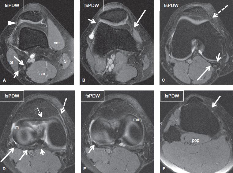
Articular surface of patella and femur, condyle, epicondyle and muscles (popliteus, sartorius, gastrocnemius, semimembranous with tendos.) the images obtained were exported to jpeg from dicom data stored on the pacs (picture archiving and communicating system). Injuries such as anterior cruciate ligament, meniscus and rotator cuff tears are all easily diagnosed when there is a firm understanding and knowledge of human anatomy. Superiorly, it extends to the level of the crossing of the biceps femoris tendon, and remains superficial to fcl in this location.10 Blood supply a large portion of the levator scapulae muscle is vascularized by two branches of the thyrocervical trunk ; Both the pronounced accuracy of the mri and the high prevalence of knee disorders, makes the knee mri the most frequently ordered imaging procedure of the musculoskeletal system. T2w axial fat sat 1. Routine protocol and then assess. Please email baodo at stanford.edu While a detailed explanation of mri protocols and mr physics is beyond the scope of this text, fast spin echo (fse) mri is most commonly utilized for mri of the knee. Mr arthrogram knee loose osteochondral lesion. Mri knee anatomy knee sagittal anatomy free cross sectional anatomy mri knee mri diagnostic imaging : The deepest layer consists of the popliteus muscle and its tendon passing. To realign the anterior cruciate ligament parallel with the sagittal imaging plane.
Mri knee anatomy scroll using the mouse wheel or the arrows. Use the mouse scroll wheel to move the images up and down alternatively use the tiny arrows (>>) on both side of the image to move the images. When interpreting the proton density images it. Intensity corresponds to a pathologic lesion. Medical images from an mri allow medical professionals to distinguish body tissues, including the meniscus (shock absorbers in the knee), cartilage, tendons, and ligaments.
Epos Trade from epos.myesr.org Saddle joint between patella and femur; Two condylar joints between femur and tibia; The deepest layer consists of the popliteus muscle and its tendon passing. Radiology knee this app is a valuable tool for radiologists surgeons medical students doctors and nurses. Anatomy of the ankle and foot in mri. Mr imaging of knees having isolated and combined ligament injuries. When a muscle has different orientations of the tendons it means that there are different patterns of edema possible depending on the tendon injured. Mri for evaluating knee pain in older patients:
Blood supply a large portion of the levator scapulae muscle is vascularized by two branches of the thyrocervical trunk ;
Knee muscle anatomy mri while a detailed explanation of mri protocols and mr physics is beyond the scope of this text, fast spin echo (fse) mri is most commonly utilized for mri of the knee. This article is based on a presentation given by david rubin and adapted for the radiology assistant by robin smithuis. In one investigation, depicted only on the proton density weighted images. View of the anatomical labels. This represents an accessory ossicle found in 10 30 of the normal population. Knee muscle anatomy mri : Sagittal pd (te<30) and fat sat pd (te36) fov14, slice 3mm. Both the pronounced accuracy of the mri and the high prevalence of knee disorders, makes the knee mri the most frequently ordered imaging procedure of the. Main supply are the genicular branches of the popliteal artery; Superiorly, it extends to the level of the crossing of the biceps femoris tendon, and remains superficial to fcl in this location.10 Use the mouse scroll wheel to move the images up and down alternatively use the tiny arrows (>>) on both side of the image to move the images. Doctors may recommend a knee mri if a patient experiences the following(3): Anatomy basic knee mri checklist.


0 Komentar